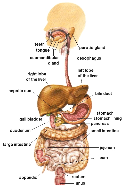
Digestive System
The Alimentary Tract
Mouth/Buccal Cavity
This is the first part of the alimentary canal and is responsible for the initial processing of ingested food. It is the only part with a bony skeleton (i.e. the jaws and teeth) and is lined with wear and tear, stratified squamous epithelium.
Enclosed in the cavity is the tongue, a muscular structure which has three main functions:
- break up food to aid mastication.
- aid swallowing.
- taste.
Chewing food stimulates the production of saliva (approximately 1.5 litres per day) which lubricates the food and also contains the enzyme salivary amylase, responsible for the initial breakdown of the large polysaccharides Starch and Glycogen into Maltose.
Oesophagus
Swallowing is an initially voluntary process that becomes a reflex reaction once food has come in contact with the pharynx. The bolus is directed across the laryngeal orifice by the epiglottis (the tracheal circular muscle also constricts) and is passed to the gut by peristalsis.
Stomach
The most dilated part of the alimentary canal and can comfortably accommodate up to 1.5 litres of food. It is contained by an elastic walled membrane and can therefore stretch even further although it can now be felt and become uncomfortable.
The functions of the stomach are:
- initial digestion of protein.
- store of food.
- churning of food.
- antibacterial device.
- absorption of water, alcohol and some drugs.
Food plus saliva and salivary enzymes enter the stomach through the cardiac sphincter. Some salivary enzymes are immediately deactivated by the low pH but many are protected from this initially by the food bolus and may continue to work for 30 minutes after swallowing resulting in up to 75% of starch breakdown.
The food now undergoes a physical and chemical process of digestion that prepares it for the small intestine. The first stage of this involves the mechanical liquefaction of the food, which also mixes the food with the digestive enzymes. This is achieved by rhythmic peristaltic waves once every approximately 20 seconds and produces a semi-fluid called chyme.
Stomach Secretions
Chemical digestion in the stomach primarily breaks down proteins into polypeptides (peptones) achieved by the secretion of the enzyme pepsin. The problem is that a protein-digesting enzyme could not exist inside a cell because it would digest the cellular contents. The enzyme existing in an inactive form called pepsinogen overcomes this. Pepsinogen is activated by hydrochloric acid secreted by different cells.
The active enzyme pepsin should now digest the wall of the stomach? This is avoided by a layer of mucus (polysaccharide) secreted by yet other cells in the stomach wall. This provides a barrier between the contents of the stomach and the stomach wall and also binds the food together and lubricate it for ease of transport through the gut.
After a period of time, depending on the nature of the food, the stomach contents are discharged through the pylorus (pyloric sphincter) into the small intestine.
Duodenum - main site of digestion
This is the first part of the small intestine. Peristalsis continues to occur to move the food along the gut (not primarily concerned with mechanical processing of food). Enzymes discharged into the lumen of the duodenum are responsible for the digestion of polypeptides, carbohydrates, lipids and nucleic acids. The pancreas, a large gland lying just below the stomach, plays a very significant role here.
Pancreatic secretions
- Water - A lot of water that has been absorbed into the blood from the stomach is replaced here.
- Sodium Hydrogen Carbonate - helps neutralise the stomach acid and provides a suitable pH for the pancreatic enzymes.
- Amylase - breaks down starch into maltose.
- Lipase - breaks down lipids into fatty acids and glycerol.
- Nucleases - breaks down nucleic acid into nucleotides.
- Protease - breaks down the polypeptides produced by the stomach into peptides, in this case, the enzyme trypsin which is again in an inactive form trypsinogen.
Duodenal secretions
- Enterokinase - activates trypsinogen into trypsin, i.e. similar role to HCl in the stomach.
- Mucus - secreted by goblet cells, provides the same role as in the stomach.
- Sodium hydrogen carbonate - this is for the initial neutralisation of the stomach acid.
Liver secretions
- Bile - This is a green watery alkaline fluid containing cholesterol, bile pigments and sodium and potassium salts of the so-called bile acids. It is produced continually in the liver approximately 5000-1,000 cm3 per day and passes through a series of ducts to the gall bladder where it is stored (approximately 30 cms3). Water re-absorption here concentrates the bile. The bile is pumped into the duodenum where it is involved in the digestion of fats. The bile salts emulsify the fats thus increasing the surface areas so that lipase digestion is more rapid.
N.B. Bile does NOT digest the fats.
Ileum - main site of absorption
The duodenum/ileum shows a number of structural and physiological adaptations to facilitate effective absorption of the now much smaller molecules. The most important gross structural modification is concerned with increasing the surface area of the gut.
- Fold - the gut is folded and this increases the intestinal surface area by a factor of about 3.
- Villi - these massively increase the surface area, They contain an abundant network of blood capillaries and an extensive lymphatic network. They also contain fine strands of smooth muscle which facilitates blood and lymph flow.
- Microvilli - this produces the brush border which vastly increases the surface area of the villi. Each microvillus is approximately 1µm high and 0.1µm in diameter. There are 2-3,000 on every epithelial cell providing final surface area for absorption of about 2 million cm2.
Process of absorption
The process of absorption occurs through the absorptive cells on the villus epithelium. Food molecules pass through these cells by phagocytosis and pinocytosis.
The micorvilli posses a wide range of specific enzymes (e.g. maltase, sucrase, lactase) to digest a wide range of molecules. They also posses active transport systems to rapidly transport the more useful food molecules into the cell cytoplasm where they are further modified by other enzymes prior to moving into the blood vessel of the villus. Less important food molecules are absorbed passively and therefore much more slowly.
Lipids are absorbed as glycerol and fatty acids or in some cases as small monoglcerides. The small ones pass straight into the blood whereas larger fatty acids are re-synthesised into fats by the absorptive cell membranes prior to being transported away through the lacteal into the lymphatic system.
The small intestine is also responsible for the re-absorption of large amounts of water, largely from internal secretions. Nine litres pass through the small intestine every day and approximately 80% is reabsorbed here. (This is greater than the volume of the blood). Food takes about three hours to pass through the small intestine before passing into the large intestine.
Colon (large intestine)
At the end of the small intestine, the chyle passes through the ileocaceal valve into the caecum and through to the colon. By this stage digestion and absorption is complete but the intestinal fluids are still extremely fluid as a result of the water that has been pumped into the small intestine throughout the digestive process.
The prime function of the colon is to reabsorb water and thus prevent dehydration. There are also bacteria present in the colon which produce vitamin K which is essential for blood coagulation.
When the descending colon has become full, the semi-solid faeces are passed into the rectum where they are stored. The distension of the rectum brings about the urge to defecate. Faeces are approximately 75% water by dry weight. The solid component is comprised of 33% bacteria, 15% inorganic salts, 5% fats together with small quantities of mucus and dead cells. The remainder is fibre. The bile pigments bilirubin and biliverdin produce the colour.

