Histology
The basic structural and functional unit of biological organisms is the cell. Tissues are made up of cells that are similar in their structure and the study of tissues is called histology. Most practical examination of cells is undertaken using a light microsocope which can magnify up to approximately 400 times (or 1500 times using an oil immersion lens). Most cell organelles are extremely small and therefore can only be studied using an electron microscope.
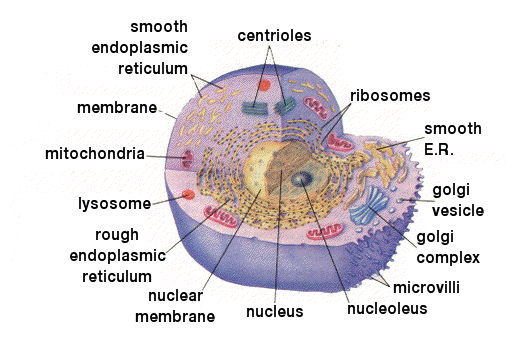
Protoplasm
This is the living material of cells differentiated into cytoplasm and nucleus.
Cytoplasm
Cytoplasm is the collective name of the aqueous ground substance plus cell organelles. The ground substance, cytosol, is a thick, semi-transparent elastic fluid containing suspended particles. It is comprised of 75-90% water together with carbohydrates, inorganic substances, proteins and lipids.
Cytoplasm is the medium in which the cell's organelles are held and many chemical reactions occur.
Nucleus
The nucleus controls cell function (including division) and stores genetic code.
The largest cell organelle, it is easily seen under the light microscope. It consists primarily of dense, gel-like nucleoplasm.
The genetic material of the cell, consisting principally of DNA, is found in the nucleoplasm. Prior to and during cell division the DNA shortens and coils into rod-shaped bodies called chromosomes. The chromosome is made of genes, each one being a strand of DNA - the molecule that stores genetic information.
The nucleus is separated from the cytoplasm by a double membrane called the nuclear membrane or nuclear envelope. Minute pores in this membrane allow the nucleus to communicate with the E.R. (endoplasmic reticulum).
Nucleoli
The nucleoli is responsible for sythesising ribosomes. These are even more dense, discrete organelles within the nucleus, comprised mainly of RNA with some DNA and protein. They are involved directly with the synthesis of protein and ribosomes which are transported to the E.R.
E.R. (endoplasmic reticulum)
The E.R. is a network of interconnecting flattened channels or cavities, which are generally continuous with the nuclear membrane. It forms a framework upon which the rest of the cell is built and therefore acts as a cell 'skeleton'. However, its principle function is as a transport system for large, and generally insoluble molecules. The E.R. is also responsible for transporting substances out of the cell.
Rough E.R.
Most of the E.R. found in cells is encrusted with ribosomes and is called rough endoplasmic reticulum. It is on these ribosomes that proteins are synthesised and then transported by the E.R.
Newly synthesised proteins are often modified as they pass along the E.R. Rough E.R. is abundant in protein secreting cells, e.g. the pancreas.
Ribosomes
Ribosomes are the site of protein synthesis and function as factories under the control os nucleic acid.
Ribosomes usually aggregate in long chains (5-50) called polyribosomes or polysomes and their presence throughout the E.R. produces a rough appearance, thus the term rough endoplasmic reticulum.
Polyribosomes are responsible for the synthesis of extracellular proteins which are principally enzymes. Unattached ribosomes are scattered in the cytoplasm and these are responsible for the synthesis of intracellular protein.
Smooth Endoplasmic Reticulum
Smooth E.R. refers to the endoplasmic reticulum not encrusted with ribosomes. It is not continuous with rough E.R. and is functionally different. It is tubular rather than flattened and is concerned with the synthesis and transport of lipids and steroids, and is therefore abundant in steroid producing cells, e.g. the adrenal cortex.
Golgi body
This is a region of particularly dense cytoplasm consisting of 4-8 flattened sac like channels. Its function is to conjugate proteins with a carbohydrate to produce glycoprotein.
These cell secretions are then enveloped and 'pinched off' to produce vesicles which move to the edge of the cell where they are secreted (exocytosis). Lysosomes are also synthesised in this way.
Lysosomes
These are small, spherical , dark structures derived from Golgi complexes and containing powerful enzymes responsible for lysis or splitting of chemicals. Their function is threefold:
- digestion of food particles in and on the membrane surface.
- destruction of old an worn out organelles.
- when a cell is damaged, the lysosomes release their enzymes to facilitate autolusis.
Lysosomes are also extremely important in bone reshaping during growth.
Mitrochondria
Spherical or rod shaped structures appearing throughout the cytoplasm.
Mitochondria are the site of cellular aerobic respiration and thus where energy is liberated for the cell's metabolic requirements in the form of ATP.
A metabolically active cell, e.g. muscle, will have a numerous large mitochondria whereas a relatively inactive one, e.g. epithelium, will have comparatively fewer and smaller mirochondria. A typical cell contains approximately 1,000 mitochondria.
Centrioles
A pair of centrioles arranged at right angles to each other is present in most animal cells. Each one is a small hollow cylinder comprised of a ring on 9 evenly spaced bundles.
They are important during cell division when they act as centres about which the microtubules involved in chromosome movement (spindle) are organised.
Cilia
Cilia posess the ability to bring about movement of a particle over a specific surface, e.g. the trachea..