The Brain and Spinal Cord
Structure of the Brain
The brain is the organ that fills the cranial cavity and is responsible for control of the body and mind. it is made up of:
- Cerebrum - two hemispheres
- Cerebellum
- Brain stem
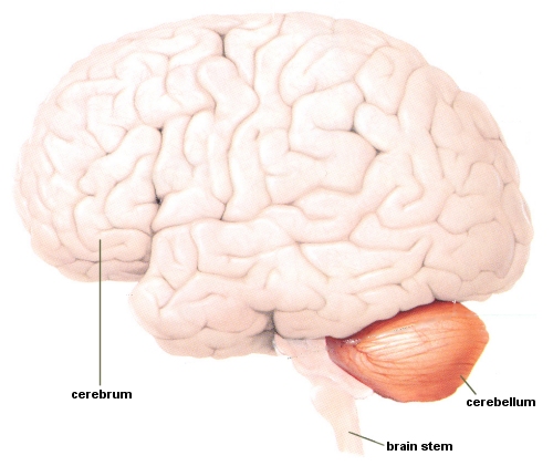
Cerebrum/Cerebral Hemispheres
The cerebrum has two hemispheres and is the largest structure of the brain. The outer layer of each hemisphere is made of folds of grey matter. Inside is made up of white matter.
The cerebrum is responsible for cognition, thinking and memory.
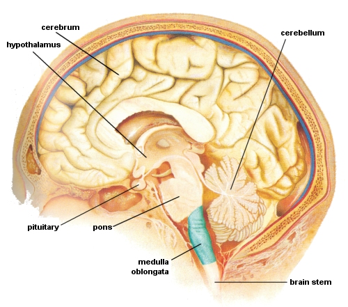
Hypothalamus
The Hypothalamus is situated at the base of the midbrain and forms the floor of this part of the brain.
The Hypothalamus is responsible for homeostasis, monitors body temperature and water balance. It is also the centre for emotions - such as pain, pleasure and thirst - and also regulates the pituitary.
Brain Stem
The brain stem is situated between the cerebrum and cerebellum and relays messages between the spinal cord and the brain.
Pons Varolii
The Pons Varolii is situated in the front of the cerebellum and consists of nerve fibres which bridge the gap between the two hemispheres of the cerebrum.
Cerebellum
The Cerebellum (also known as the small brain) is situated in the posterior cranial fossa behind the pons varolii. Like the cerebrum, it also has two hemispheres, with grey and white matter in it.
The Cerebellum is responsible for fine motor control, balance and co-ordination, reflex and conscious action and muscular posture and tone.
Medulla Oblongata
The Medulla Oblongata is the lowest part of the brain stem situated above the spinal cord. It has white matter on the surface and grey matter inside and is known as the vital centre because it controls the actions of the heart and lungs.
The Medulla Oblongata is responsible for homeostasis, breathing, heart rate, hearing, balance, posture, and the reflex centre (salivating, coughing and vomiting).
Pituitary
The Pituitary is a gland that is situated at the top of the brain stem in the frontal part of the brain.
The Pituitary is the interface between the nervous and endocrine/hormonal system.
Structure of the Spinal Cord
The spinal cord is the other part of the Central Nervous System and extends from the medulla oblongata, through the spinal vertebrae and ending at the first lumbar vertebra. It consists of white matter on the surface and grey matter in the centre.
The following groups of nerves constitute the spinal nerves:
Cervical pairs (8)
Cervical Plexus - The first 4 cervical nerves; supply the skin and muscles of the neck, shoulder plus the phrenic (diaphragm) nerve
Brachial Plexus - The lower 4 cervical and 1st thoracic; supply the skin and muscles from the neck base to the fingers
Thoracic pairs (12)
Thoracic Plexus - supplies the chest muscles and most of the abdominal wall
The remaining nerves all leave the vertebral column at the 1st lumbar vertebra and extend down in the cauda equina:
Lumbar pairs (5)
Lumbar Plexus - The first 3 and part of 4th; supplies the skin and muscles of the lower abdomen, thighs and groin
Sacral pairs (5)
Sacral Plexus - The 4th and 5th lumbar and first 3 sacral; supplies the skin and muscles of the pelvis and hamstrings and includes the sciatic nerve
Coccygeal pair (1)
Coccygeal Plexus - The last 2 sacral and coccygeal nerve; supplies the skin and muscle of the remaining pelvic area, including anal sphincter and external genitalia

