The Urinogenital System
Urinary System
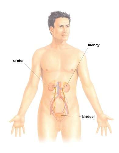
The urinary system is responsible for the removal of toxins from the body, such as alcohol and urea, through the process of filtration and excretion. It is comprised of a pair of kidneys, ureters, a bladder and a urethra.
Kidney
The human body has two kidneys, situated at the posterior wall of the abdomen in the upper lumbar part of the back, either side of the spine. During circulation, one quarter of the total blood in circulation passes through the kidneys so that toxic substances can be filtered.
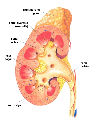
The kidney has two main parts - the cortex on the outside and the medulla on the inside. The medulla leads into the renal pelvis and in the centre is the hilum, where blood vessels, lymphatic vessels, nerves and the ureters enter the organ.
Renal Pelvis
The renal pelvis connects the medulla to the ureter. It collects urine from the tubules in the medulla and passes it to the ureter.
Ureter
The ureter acts as a conduit for urine to pass from the pelvis to the bladder.
Bladder
The bladder is situated in the pelvic cavity and acts as a reservoir for urine. When approximately one fifth of a litre of urine is present, the liquid stimulates the autonomic nerve endings in the bladder wall and the bladder contracts. An internal sphincter of the bladder subsequently relaxes and allows the urine to pass into the urethra and ultimately out of the body.
Nephron
Blood enters the kidneys via the afferent arterioles. The formation of these vessels is called the glomerulus which is surrounded by the Bowmans Capsule. The blood is under pressure and water and other substances pass through into the capsule. Proteins and blood cells remain in the blood. The Bowmans Capsule thus serves as a collection point of both waste and non-waste products. It is the subsequent phases of reabsorption that are responsible for retaining these non-waste substances.
The proximal convoluted tubules allow the filtered substances to travel into the Loop of Henle and on to the distal convoluted tubule, so that reabsorption can take place. The Loop of Henle is surrounded by blood vessels and the walls of the tubules are able to let water, glucose, salts and ions back into the blood. In fact, only about 1% of the liquid filtered into the Bowmans Capsule is excreted as urine.
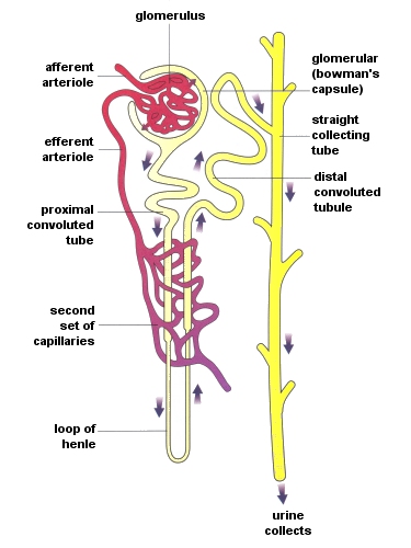
The nephron eventually straightens out and joins the collecting tubule in the medulla. These tubules form pyramids in the medulla and lead into the pelvis and subsequently the ureter.

